©1999-2007 by S.M. Gon III
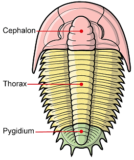
The trilobite cephalon (orange) is the most anterior of the three trilobite tagmata (major body sections). It bears eyes, mouthparts and antennae,
and articulates with a thorax (yellow) of multiple articulated segments (that in some species allowed
enrollment), and a pygidium,
(green) or tail section of fused segments.
|

The underside of the cephalon reveals the hypostome (H), a mouthpart that underlies the glabella (G). The function of the hypostome and its role in classification is discussed on a page dealing with hypostome terms.

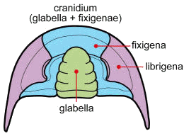
The axial portion of the cephalon is called the glabella (green), and is largely occupied by the anterior digestive system. The cheeks (genae) are the pleural lobes on each
side of the glabella. When trilobites molt or die, the
librigenae (purple) or "free cheeks" often separate along the facial sutures, leaving only the cranidium --that is, the combined glabella and fixigenae (blue) or "fixed cheeks"
|

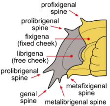
Terms for genal spines:
while the typical cephalic spine placement is at the genal angle (the
lateral posterior corner of the cephalon), where it is called simply a
genal spine, other spine locations may be
anterior (pro) or adaxial (meta) of the genal angle, and are further
defined by their placement on either the fixigena (shown in yellow
here) or the librigena (in purple). Note that a prolibrigenal spine
might occur close to the genal angle, or be placed far forward along
the anterior margin of the cephalon. A profixigenal spine is
usually on the anterior margin. In some trilobites, a series of
small spines might be present along most of the genal margin (e.g., see
specimens in the family Odontopleuridae)
|

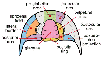
When describing
differences between different taxa of trilobites, the presence, size,
shape, and proportions of the cephalic features are often
involved. The diagram above indicates via colors some of the important
features use to distinguish taxa. For example, if the glabella is
large, it reduces the size of
the preglabellar area, in some groups displacing it altogether. To the
right are cephalic (cranidial) features
described when the librigenae are missing.
|


