Trilobite Facial Sutures
last revised 03 February 2009 by S.M. Gon III
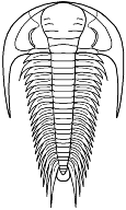
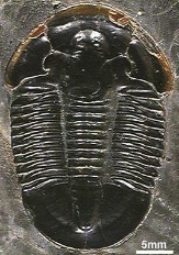
facial sutures on Asaphiscus wheeleri
The facial sutures of Asaphiscus run from the anterior of the cephalon to the eye, then to the back of the cephalon.
What are facial sutures?

They are lines on the cephalon along which the parts of the cephalon separate when the trilobite molts (sheds its exoskeleton -- see animation and discussion below). They typically run from somewhere along the anterior edge of the cephalon, toward and around the edge of the eye, and continue from there to end at either the side or rear of the cephalon. They are vital for proper molting and growth of  trilobites, and they provide us with another character with which to help classify different trilobite groups.

There are three main categories of facial suture types (proparian, gonatoparian, and opisthoparian), described below. If you want more detailed definitions of the terms related to facial sutures, they can be found in the on-line glossary of trilobite terms.
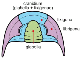
The facial sutures determine the boundary between the central cranidium (glabella and fixed cheeks) and the librigenae (free cheeks). Trilobite specimens often lack librigenae. They are lost during molting, and also disarticulate easily following the death of the trilobite. 
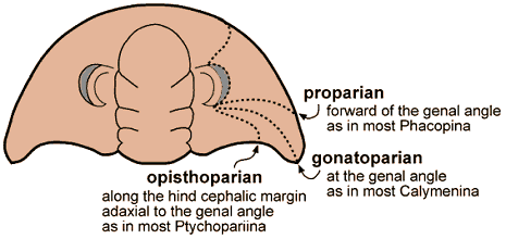
The three main types of facial sutures
are defined by where the suture ends, relative to the genal angle:

All images this page except where noted ©1999 - 2009 by S. M. Gon III. created using Freehand

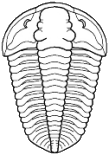
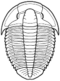
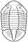
Here are some examples of the three suture types:

Eodiscina
(proparian)
Phacopina
(proparian)
Calymenina
(gonatoparian)
Ptychopariina
(opisthoparian)
Asaphida
(opisthoparian)


Suture patterns within types:
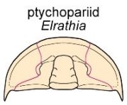
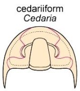
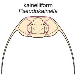
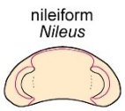
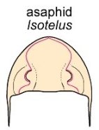
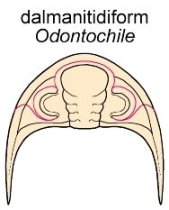
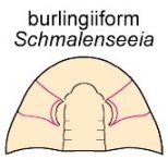
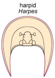
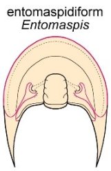
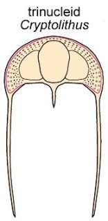
Facial sutures once played a primary role in defining family-level relationships, and several common patterns within the three types above were named according to the families that bore the suture pattern. While many of the patterns are more or less valid and useful, more recently it has been observed that suture form and pattern can vary significantly even among species of a given genus, and the primacy of facial sutures in classification has declined. The presence or absence of sutures is still used to distinguish between the earliest trilobites, with those lacking sutures placed in the suborder Olenellina of the order Redlichiida, and those bearing sutures placed in either the Redlichiina of Redlichiida, or the order Ptychopariida.  In the images below of some of these classic suture patterns, the course of the sutures is shown in red. The name of the pattern is above and the name of the trilobite genus representing the pattern is presented below in italics. Most of the patterns are variations on the opisthoparian type, but a few proparian patterns are also shown. The marginal pattern shown by harpetids and trinucleioids is a derivation of opisthoparian type (as shown by the entomaspidiform pattern).
   
Ptychopariid sutures diverge slightly from the eyes and run more or less straight to the anterior border. Cedariiform posterior sutures cross the lateral border, then swing backward and inward to meet the posterior margin. Kainelliform sutures diverge widely until they cross the anterior border, then swing strongly inward to run along the anterior margin. Nileiform sutures converge and meet before reaching the anterior margin, running parallel to the anterior border. Asaphid sutures converge and meet at an apex at or near the anterior margin, then form a median suture


Dalminitidiform sutures are proparian, with anterior sutures that converge and meet, closely following the anterior border; also called phacopid pattern. Burlingiiform sutures are proparian, with anterior and posterior sutures running subparallel to create roughly rectangular fixigenae Harpid stures are purely marginal, running along the cephalic margins; the eyes are often vestigial and no longer involved with the sutures Entomaspidiform sutures run mostly along the margin, but involve the eyes via two narrow strips.  Trinucleid sutures are also largely marginal, but may run parallel to the margin on the dorsal cephalic surface.


Sutures and Ventral Cephalic Structures
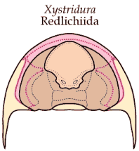
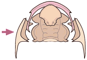
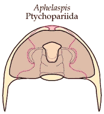
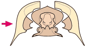
The dorsal sutures do not end at the margins, but extend under the cephalon, involving the cephalic doublure, and interacting with the rostral plate (shown pink in the images below below) and the hypostome. Because it is difficult sometimes to envision the nature of the dorsal and ventral sutures, and how they partition the cephalon, I have developed images below that show the sutures in an intact and a disarticulated cephalon of representative trilobites, including ones with large to small rostral plates, and one lacking a rostral plate. Note that in species with conterminant hypostomes (ones attached firmly to the anterior boublure), the hypostome may either have well-developed sutures, or may be permanently atached to the rostral plate.

images ©2003 by S. M. Gon III
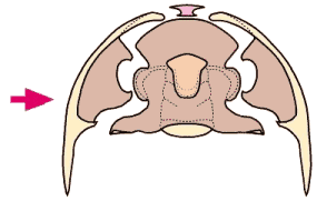
In Xystridura, a typical redlichioid trilobite, there is a large rostral plate (pink) that involves the anterior and lateral doublure. The hypostome is probably permanently attached to the doublure, so that when the sutures separate, it remains docked to the rostral plate.
In Aphelaspis, a typical ptychoparioid trilobite, there is a small rostral plate (pink), and the hypostome is natant, so is never attached to the narrow cephalic doublure, but "floats" beneath the front of the glabella (held in place by muscles and connective tissue to the glabellar apodemes). 
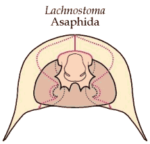
In Lachnostoma, a typical asaphoid, the free cheeks split evenly along a median ventral suture. There is no rostral plate. There is a hypostomal suture, which allows the hypostome to separate as needed. Notice that the cephalic doublure is extremely wide.

Molting (Ecdysis) and the Role of Sutures


Trilobites, as arthropods, grew by shedding their old, rigid exoskeleton in a process called ecdysis, or molting.  They underwent this process several times during their development. The animation below depicts a Paradoxides trilobite molting (left is the animal in top view). In the animation, the facial sutures (red) split, opening the cephalon (separating the cranidium from the librigenae and associated structures). This provides an exit for the molting trilobite (purple) from its old exoskeleton (orange). The arching of the body plants the rear pleural spines securely into the substrate and likewise anchors the rostral plate at the anterior cephalic margin downward, providing an exit ramp for the emerging newly-molted animal. This sequence is consistent with fossils of Paradoxides in which the librigenae and rostral-hypostomal plate are found inverted beneath the axial cranidial shield. All it would take is slightly more forward tilting to have the molted librigenal assemblage flip into such an inverted position.
Ahh! Now I can stretch a bit!
This animated image ©2000 by S. M. Gon III
Adapted from Whittington, 1990 via the 1997 Treatise (p. 156-7). 

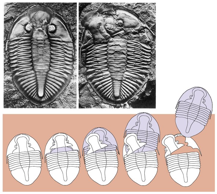
Fossil evidence of the molting sequence
Some trilobite models come complete with sunroof...
Fossil photos (top) from Ludvigsen 1979a. Molting sequence figure (bottom) adapted from Ludvigsen 1979b.
In another example of the consequence of molting, the displaced cranidium and librigenae of the Canadian trilobite Pseudogygites latimarginalis in the photographs shown below is explained by the reconstruction of the molting sequence shown under the photographs: The facial sutures separate, allowing the molting trilobite (purple) an opening through which it emerges from its old exoskeleton (white).
Sources cited:
  • Ludvigsen, Rolf.  1979a. The Ordovician trilobite Pseudogygites Kobayashi in eastern and arctic North America.  Royal Ontario Museum Life Sciences Contributions 120, 41 pages.
  • Ludvigsen, Rolf.  1979b.  Fossils of Ontario.  Part 1: The Trilobites.  Royal Ontario Museum Life Sciences miscellaneous Publications, 96 pages. 
  • Whittington, H. B. 1990. Articulation and exuviation in Cambrian trilobites. Philosophical Transactions of the Royal Society of London (series B) 324:111-47. fig. 1-54.

  • stroll with me among the topics below...
    Walking Trilobite animation ©2000 by S. M. Gon III