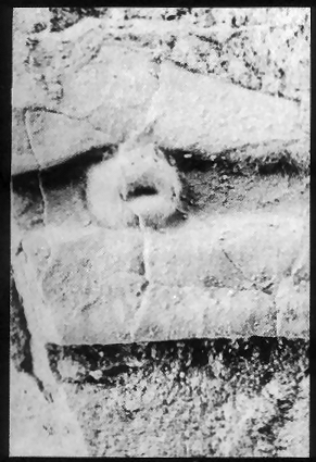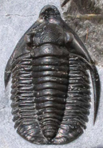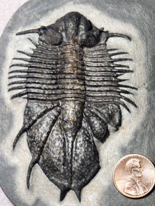
An abnormality in the pygidium of the Moroccan lichid trilobite Acanthopyge |
Introduction Because trilobites are relatively common in the fossil record, occasionally we find various kinds of abnormalities, such as asymmetrical features, healed injuries, or signs of disease. Teratology is the study of malformations or serious deviations from normal form or development, and such studies of trilobite fossils have revealed some very interesting abnormal forms. Healed injuries The most common types of trilobite abnormalities are partially healed injuries. Trilobites were victims of many predators in Paleozoic seas. Because an exoskeleton can not heal until molting, abnormalities such as the ones shown here document that the trilobite survived the attack and began to heal the damaged area during its next molt. This kind of repair would often require several molts, with more and more recovery of the injured area restored, so the molts provide a sequential picture of how trilobites repaired their wounds. |
Partial healing of two thoracic pleurae in Phacops.The rounded edges of the small pleurae indicate the zone of healing. a fresh injury would likely be sharp-edged. Image courtesy: Marc Behrendt See more at the Bedrock Bugs Gallery |
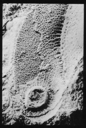
Disease-related abnormalities Disease is another possible cause of damage to trilobite exoskeletons. Infections of small injuries might cause slow-healing ulcers that induce the exoskeletal equivalent of scar tissue or even neoplasms (tumors) such as shown in the exoskeletons of Bohemoharpes and Hydrocephalus at right (from Owen 1983). In the Bohemoharpes (left), the abnormality is also accompanied by radiating circulatory canals, similar to the way that tumors often attract blood vessel development. The neoplasm in the exoskeleton of Hydrocephalus (right) may have begun as an irritation or small injury in the sensitive thinner exoskeleton between the segments. In both cases, over sequential molts, scar tissue or thickened exoskeleton was deposited around the infection site, perhaps in a similar way that tree bark surrounds and encloses injury. Typically there is only a single neoplasm on a trilobite exoskeleton. This suggests that trilobites were resistant to such infections, or that major infections proved fatal, with no opportunity for exoskeletal evidence. |
Neoplasm in the fringe of Bohemoharpes. |
Neoplasm in the interpleural axis of Hydrocephalus. |
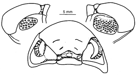
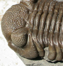
Asymmetrical development Occasionally there are specimens that show asymmetries that are relatively subtle, such as different numbers of lenses between right and left eye, where there is no evidence of injury. Such abnormalities might have arisen as a result of developmental problems. For example, in a specimen of Phacops rana (right), the size and number of lenses in the left eye is much reduced compared to the right, which bears a normal size eye and a full complement of lenses. The pattern of reduction, with smaller eye surface and reduced numbers of lenses of normal size, mimicks evolutionary reduction of eyes in other phacopids, but exhibits it in a single abnormal individual. The fact that this is a holaspid suggests that even with a smaller eye and reduced lenses, the growing trilobite was able to engage in a normal enough lifestyle to survive and grow to adult stage. |
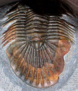
abnormal right pygidium Scabriscutellum sp. image courtesy Dieter Holland |
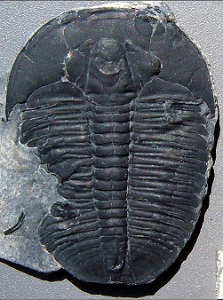
abnormal left thoracic pleurae Elrathia kingi image courtesy Ross Wetzel |
abnormally short genal spine Diademaproetus sp. image courtesy Dieter Holland |